Why Choose Anatomical Specimens for Medical Training?
Medical training demands more than diagrams, screens, and simulated models. An Anatomical Specimen gives learners direct experience with human structure, proportion, and natural variation. Students can examine tissue planes, trace vessel branches, and observe how organs relate within limited spaces. The lesson becomes tangible. That matters.
In accredited medical programs, supervised specimen-based teaching supports anatomy, surgery, pathology, and clinical communication. Experienced instructors can explain landmarks while students handle instruments carefully and follow established laboratory procedures. This approach can strengthen spatial understanding and improve confidence before clinical practice. It also encourages precise observation, respectful teamwork, and informed discussion. However, an Anatomical Specimen is not a perfect learning tool. Preservation may alter color, texture, or flexibility. Individual specimens cannot represent every patient. Some learners may also experience discomfort, even after careful preparation. That response deserves attention, not dismissal.
Ethical sourcing, documented consent, institutional oversight, and safe handling remain essential. Reliable training requires more than access to specimens. It requires qualified educators, appropriate facilities, and clear learning objectives. Published anatomical references and clinical guidelines should support laboratory observations. Educators must also acknowledge uncertainty when structures differ from textbook examples. This honest comparison develops better judgment. It reminds students that real bodies are complex, variable, and never fully predictable. Careful reflection can turn a challenging laboratory session into a lasting professional lesson.
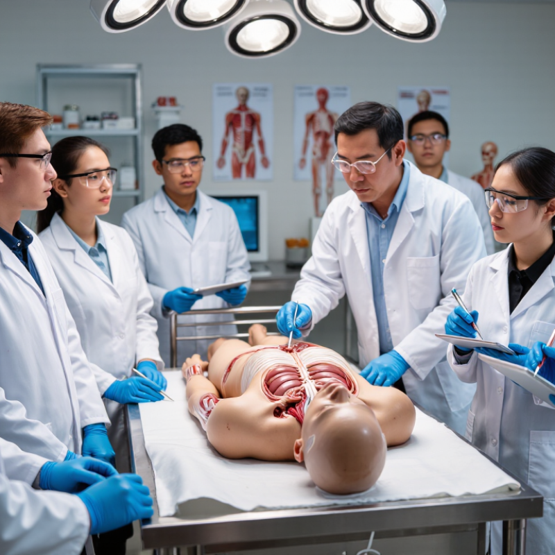
Anatomical specimens are preserved human or animal tissues used to teach body structure. They range from complete donated bodies and dissected regions to individual organs, bones, and thin tissue sections. Each format reveals different details. A skeleton makes joint relationships easy to see, while a preserved heart shows chambers and major vessels in three dimensions.
Specimens may come from documented body-donation programs or approved educational and clinical collections. Responsible programs record provenance, obtain appropriate consent, and follow institutional handling rules. Students and instructors should be able to ask where a specimen came from and how it may be used. That paperwork matters as much as the labels on a storage jar.
Training roles vary. Learners can trace nerves, compare organ shapes, and examine tissue planes that diagrams flatten. Wet-preserved specimens often retain useful texture; plastinated sections are easier to handle and store. Preserved tissue is not living tissue. It cannot show normal movement, circulation, or healing, so it works best alongside imaging, models, and supervised practice. Even a carefully prepared specimen leaves gaps. That limitation is worth discussing, not hiding.
| Specimen Type | What It Is | Typical Source | Training Roles | Key Considerations |
|---|---|---|---|---|
| Embalmed whole-body specimen | A donated human body preserved with embalming fluids for extended study. | Human body donation programs operating under applicable laws, consent requirements, and institutional policies. | Regional anatomy, dissection, anatomical variation, and foundational medical education. | Suitable for repeated study, but preservation can change tissue color, texture, and flexibility compared with living tissue. |
| Fresh-frozen human specimen | Human tissue or a body preserved by freezing rather than conventional embalming. | Consent-based anatomical donation programs and appropriately governed tissue repositories. | Surgical approaches, procedural practice, and study of tissue planes and joint movement. | Often retains more lifelike tissue properties than embalmed material; requires controlled storage, handling, and biosafety procedures. |
| Prosection | A specimen that has been dissected in advance to expose particular structures. | Prepared from donated human bodies under institutional authorization and donor-consent conditions. | Teaching anatomy when course time is limited; demonstrating complex structures and relationships. | Provides a focused view without requiring learners to perform the initial dissection; does not provide the same hands-on dissection experience. |
| Plastinated specimen | An anatomical specimen in which much of the water and fat has been replaced with a durable polymer. | Prepared from donated human or animal material by specialized facilities following relevant consent and oversight requirements. | Repeated classroom handling and close examination of organs, regions, or anatomical structures. | Durable and generally dry to handle, but does not reproduce the feel or movement of living tissue. |
| Skeletal specimen | Whole or partial bones, including articulated or disarticulated skeletal material. | Documented anatomical collections, donated material, or ethically sourced educational collections. | Bone identification, joint anatomy, landmarks, and the relationship between skeletal structures. | Useful for durable, repeated study; soft tissues, circulation, and organ relationships are not represented. |
| Pathology or surgical specimen | Human tissue removed or collected for clinical diagnosis or treatment and made available for education under appropriate controls. | Clinical care and pathology services, subject to applicable consent, privacy, legal, and institutional requirements. | Teaching gross pathology, disease-related structural changes, and the appearance of specific tissues. | Availability and educational use depend on clinical governance; patient confidentiality and proper handling are essential. |
| Animal anatomical specimen | Whole or partial animal material used to study anatomy or practice selected procedures. | Ethically and legally sourced animal material, subject to applicable animal-welfare rules and institutional review. | Comparative anatomy and selected skills training where animal anatomy is relevant to the learning objective. | Anatomy can differ from human anatomy; use should be justified by educational needs and governed by relevant welfare standards. |
Training note: Specimen selection should match the learning objective. Human anatomical material should be handled respectfully and used in accordance with donor consent, applicable law, institutional policy, and appropriate health-and-safety procedures.